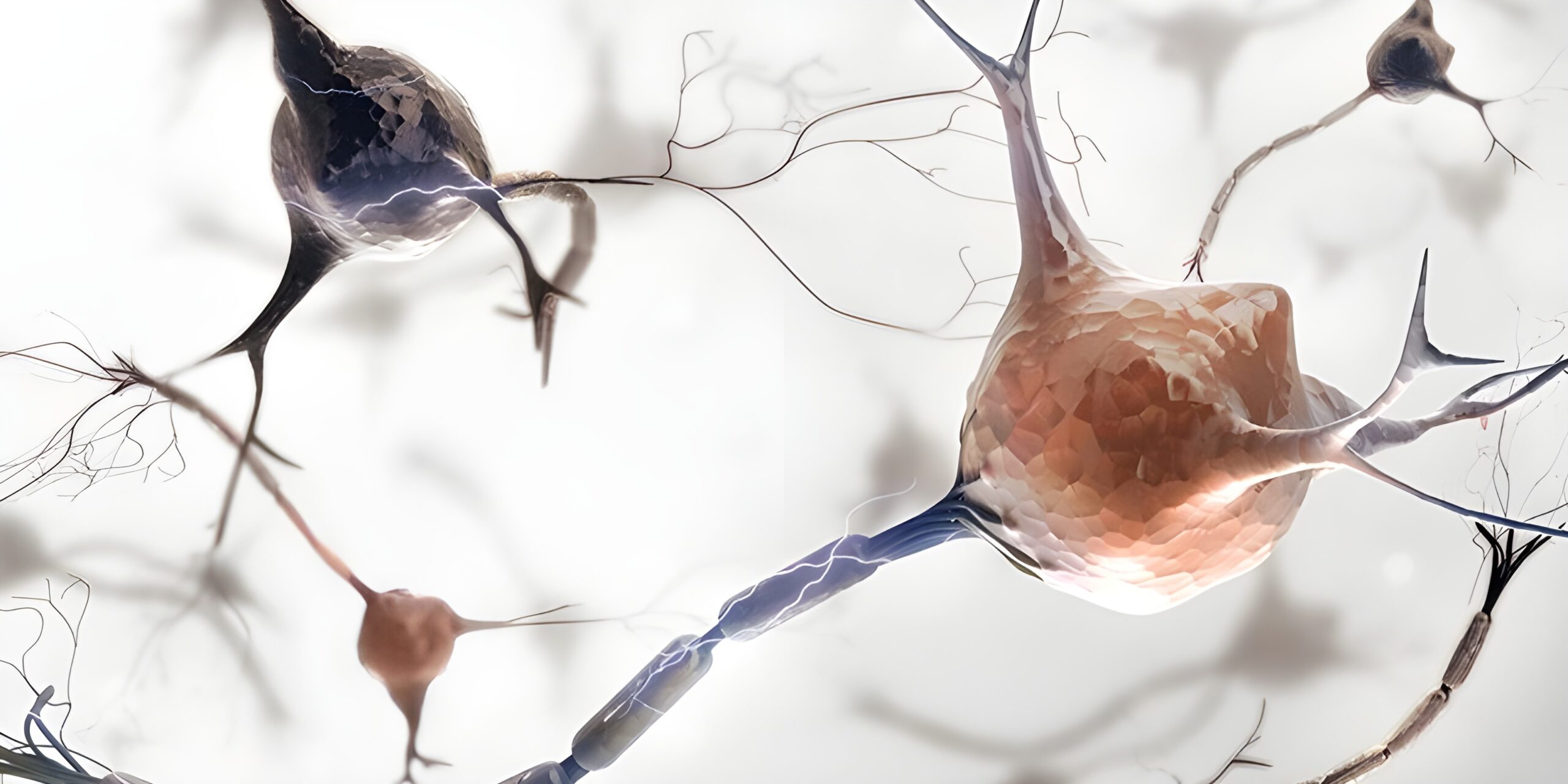
A scientific analysis with particular reference to EEG, ERP and microstate biomarkers
Summary
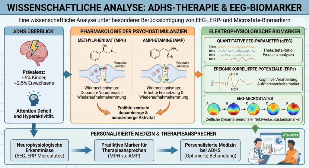
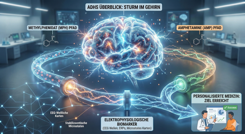
Attention deficit hyperactivity disorder (ADHD) is one of the most common neurodevelopmental disorders, affecting around 5% of children and 2.5% of adults worldwide. Psychostimulants, in particular methylphenidate (MPH) and amphetamines (AMP), represent the first-line pharmacological treatment. Although both classes of substances increase central dopaminergic and noradrenergic activity, they differ fundamentally in their mechanisms of action. This paper analyses the pharmacological differences between the two classes and their implications for clinical use. A particular focus is placed on electrophysiological biomarkers, including quantitative EEG parameters, event-related potentials (ERPs) and EEG microstates, which could serve as predictive markers of treatment response. Integrating these neurophysiological findings into clinical practice could be an important step towards personalised medicine in ADHD.
1. Introduction
Attention deficit hyperactivity disorder (ADHD) is one of the most thoroughly researched neurological conditions in modern medicine. It manifests through persistent patterns of inattention, hyperactivity and impulsivity that impair academic, occupational and social functioning. Worldwide prevalence is estimated at around 5.29% in children and adolescents and 2.5–4.4% in adults. Annual societal costs in the USA alone are estimated at USD 143–266 billion.
The pharmacological treatment of ADHD relies primarily on two classes of psychostimulants: methylphenidate-based preparations and amphetamine derivatives. Although both classes have been used successfully for decades and show a response rate of around 70%, a significant subgroup of patients responds differently to the various stimulants. This observation suggests that, despite similar clinical efficacy, fundamental pharmacological and neurophysiological differences exist.
Identifying reliable biomarkers to predict individual treatment response is an important step towards personalised medicine. Electrophysiological methods, in particular the electroencephalogram (EEG) and event-related potentials (ERPs), offer promising possibilities for characterising neurophysiological subgroups and predicting treatment success.
2. Pharmacological mechanisms of action
2.1 Common principles of action
The primary pharmacological effect of both methylphenidate and amphetamines is to increase central dopamine and noradrenaline activity. These catecholamines play a central role in the executive and attentional functions of the prefrontal cortex. The aetiology of ADHD is complex and involves multiple genetic and non-genetic factors. Current evidence, however, converges on the assumption that catecholamine neurotransmission is impaired in the brains of patients with ADHD.
Under normal circumstances, the prefrontal cortex regulates attention, behaviour and emotions. Deficits in prefrontal cortex function have been linked to ADHD symptoms such as poor impulse control, limited sustained attention and increased distractibility. Both stimulants and the non-stimulant atomoxetine increase synaptic catecholamine concentrations in the brain, particularly in the prefrontal cortex.
2.2 Specific mechanisms of action of methylphenidate
Methylphenidate acts primarily as a dopamine and noradrenaline reuptake inhibitor. Its mechanism of action is based on blocking the presynaptic dopamine and noradrenaline transporters, which leads to increased availability of these neurotransmitters in the synaptic cleft. Unlike amphetamines, methylphenidate does not promote the release of dopamine from synaptic vesicles.
Additional mechanisms of action of methylphenidate include agonist activity at the serotonin 1A receptor and a redistribution of the vesicular monoamine transporter 2 (VMAT-2). There is also evidence of interactions with glutamate and opioid systems. These mechanisms may contribute to its specific clinical effects and side-effect profiles.
2.3 Specific mechanisms of action of amphetamines
Amphetamines show a more complex pharmacological profile than methylphenidate. In addition to inhibiting the dopamine and noradrenaline transporters, amphetamines also inhibit the vesicular monoamine transporter 2 (VMAT-2) and monoamine oxidase activity. The decisive difference from methylphenidate lies in the ability of amphetamines to actively release catecholamines from presynaptic nerve terminals.
Amphetamine exists as two stereoisomers with different activity profiles. The l-enantiomer (levoamphetamine) produces more cardiovascular and peripheral effects than the d-enantiomer (dextroamphetamine). At low doses, levoamphetamine produces stronger arousal than dextroamphetamine and acts primarily on noradrenaline. At higher doses, dextroamphetamine shows stimulant properties three to four times stronger than those of levoamphetamine and acts primarily on dopamine.
2.4 Clinical implications of the pharmacological differences
The different mechanisms of action have clinically relevant consequences. Amphetamines lead to a stronger release of dopamine, which can be described as “flooding” the synaptic cleft with dopamine. Methylphenidate, by contrast, brings about a subtler, more indirect increase in dopamine availability by blocking reuptake.
These differences may explain why individual patients respond differently. Studies have shown that of 174 patients in six crossover studies, 48 responded better to amphetamines, 27 better to methylphenidate and at least 72 to both substances. This yields an overall response rate of over 87% when both classes are tried sequentially. A patient who does not respond to one stimulant should therefore try the other.
3. Quantitative EEG biomarkers in ADHD
3.1 The theta/beta ratio
The theta/beta ratio (TBR) was historically one of the most frequently studied EEG parameters in ADHD. Numerous studies reported that ADHD is characterised by increased theta activity and reduced beta activity, leading to an elevated TBR. In a multicentre study, the TBR distinguished ADHD patients from controls with a sensitivity of 86–90% and a specificity of 94–98%.
A comprehensive meta-analysis of nine studies including a total of 1,253 children and adolescents with ADHD and 517 controls found, however, that the TBR cannot be considered a reliable diagnostic marker for ADHD. The mean effect size was 0.75 for 6- to 13-year-olds and 0.62 for 6- to 18-year-olds, with significant heterogeneity between studies.
More recent research has shown that elevated TBR values in ADHD can partly be explained by slowed alpha peak frequencies, which makes the TBR a non-specific measure combining different neurophysiological subgroups. Nevertheless, the TBR retains prognostic value: elevated baseline theta and a high TBR are associated with a good response to stimulants.
3.2 Alpha peak frequency as a predictor
Individual alpha peak frequency (APF) has proven to be a promising biomarker for predicting treatment response. A low APF has been identified as a biomarker associated with non-response to stimulants in male ADHD patients. Interestingly, a low APF is also associated with non-response to antidepressants, suggesting that it could serve as a general biomarker of non-response and help in developing new treatments for such subgroups.
The iSPOT-A study (International Study to Predict Optimized Treatment for ADHD), a multicentre, international, prospective open-label study of 336 children and adolescents with ADHD, examined TBR and APF as predictors of methylphenidate response. 62% of the ADHD group were classified as responders. Responders did not differ from non-responders in age, medication dose or baseline severity of ADHD symptoms.
3.3 EEG spectral power in adults
For adult ADHD patients, studies show that EEG spectral power, but not the TBR, can function as a neuromarker. A machine-learning classification analysis found that the best predictors of ADHD status were increased power in delta, theta and low alpha over centro-parietal regions, as well as in frontal low beta and parietal mid beta.
In adults with ADHD, consistently increased absolute and relative theta power and alpha activity were found compared with controls, together with a general reduction in neural activity during tasks requiring attention. No consistent evidence of an atypical TBR was found in adult ADHD, suggesting that the TBR may be a characteristic of childhood ADHD.
4. Event-related potentials (ERPs)
4.1 The P300 component in ADHD
The P300 (or P3) is an ERP component that reflects attentional and memory processes. It can be divided into P3a and P3b subcomponents. A recent meta-analysis of 52 articles concluded that group differences between ADHD and control participants were most robust for late ERPs. The ADHD group showed smaller cue P300 amplitudes, longer Go P300 latencies and smaller NoGo P300 amplitudes than the non-ADHD group.
The P3a is regarded as a reflection of frontal lobe function and occurs when attention is shifted from frequent target stimuli to novel or rare stimuli. It is linked to early cognitive processes such as attentional mechanisms and working memory and is influenced by dopaminergic activity. The P3b, on the other hand, reflects later processing in temporo-parietal regions and is associated with memory updating and storage.
4.2 Effects of methylphenidate on ERPs
A systematic review and meta-analysis of methylphenidate effects on the P300 in ADHD found that, when comparing pre- and post-MPH effects in the same individuals, P300 amplitude tends to increase after administration in central and posterior regions. A moderator analysis by task type showed a larger effect size for Go/No-Go tasks.
In a study of 26 boys with ADHD, EEG results after 8 weeks of methylphenidate treatment showed a reduction in NoGo P300 latency at electrodes Fz, Cz and Pz compared with baseline. Normalisation of P300 latency after MPH treatment appears to be a reliable neural biomarker of a positive treatment response in children with ADHD. MPH was associated with improvements in executive functions, particularly inhibitory control and working memory.
4.3 P300 topography as a predictor of treatment response
Auditory P300 topography can predict response to stimulants in ADHD patients. Patients with a right fronto-central to parietal auditory P300 amplitude ratio greater than 0.5 respond robustly to stimulants. One study confirmed that P300 amplitude at electrode T8 can predict response to methylphenidate, with a value greater than 7.65 µV predicting response.
The mean 31-electrode P300 amplitude was able to predict response to atomoxetine. Different P300 parameters could thus help predict whether a stimulant or a medication with a different mechanism of action should be used. In a study of 87 paediatric ADHD patients, an ERP-based index for predicting medication response distinguished significantly between responders and non-responders, with a large and clinically meaningful effect size of 1.84.
4.4 Differential ERP markers for methylphenidate vs. amphetamines
Ogrim and colleagues have repeatedly found that attenuated pre-treatment P3 amplitudes during a cued Go/No-Go task predict a positive behavioural response to methylphenidate or dexamphetamine stimulants. ADHD symptoms associated with atypical patterns of intrinsic neural activity may be less responsive to methylphenidate. In contrast, ADHD symptoms associated with abnormal fronto-striatal network arousal may be correctable with methylphenidate.
Investigating the EEG correlates of methylphenidate response in ADHD showed that medication administration led to increased alpha activity in central and parietal regions. Children who responded to the medication showed increased frontal beta activity, whereas non-responders showed decreased beta activity in the same region. Increased frontal beta activity was significantly correlated with medication-related improvements in sustained attention and response inhibition.
5. EEG microstates as functional biomarkers
5.1 Basics of microstate analysis
EEG microstates represent an alternative approach to studying large-scale cortical dynamics in ADHD. Microstate analysis models the spontaneous EEG as a sequence of states defined by the recurring occurrence of particular distributions of scalp potentials. These different states remain stable for about 100 milliseconds and switch from one to another.
Neuroscientists typically distinguish five main microstates (A to E), which manifest different spatial configurations in the electrical field of the EEG. The microstate technique considers the combined spatial and temporal aspects of brain activity and has the advantage of using the full spatial resolution of high-density EEG recordings.
5.2 Microstate alterations in ADHD
A landmark study by Férat and colleagues investigated EEG microstates as novel functional biomarkers for adult ADHD using two independent datasets. Spatially, ADHD and control participants showed equivalent microstate topographies (canonical maps), indicating that prototypical EEG generators are preserved in ADHD. This agreement, however, was accompanied by significant differences in temporal dynamics.
At group level and across both datasets, an ADHD diagnosis was associated with longer mean durations of a fronto-central topography (microstate D). This suggests that its electrocortical generators could act as pronounced “attractors” of global cortical dynamics. In addition, one of the datasets showed evidence of reduced time coverage and mean duration of microstate A, which correlated inversely with ADHD scores, while microstate D metrics correlated with sleep disturbances.
5.3 Microstate D and attentional functions
Microstate D has repeatedly been linked to attentional functions and to brain regions in dorsal attention networks. ADHD patients often show an excess of microstate D, a state characterised by fronto-central cortical activity. A recent review of 13 studies applying microstate analyses to resting-state and task-based brain activity in ADHD populations consistently confirmed changes in microstate organisation, with increased duration (MS-D) and changes in temporal dynamics (MS-C).
These changes may reflect executive dysfunction and delayed maturation of the default mode network. Microstate B showed promising potential for distinguishing between ADHD subtypes based on differences in visual network function. Task-based and ERP studies using paradigms such as the Continuous Performance Task or Go/No-Go task identified microstate abnormalities linked to inhibition and the allocation of attentional resources.
6. Clinical effectiveness and network meta-analyses
6.1 Comparative efficacy in children and adolescents
The most comprehensive network meta-analysis of the comparative efficacy and tolerability of ADHD medications analysed double-blind, randomised controlled trials of amphetamines, atomoxetine, bupropion, clonidine, guanfacine, methylphenidate and modafinil. In children and adolescents, based on clinician ratings, all medications were better than placebo.
In head-to-head comparisons, differences were found only in efficacy (clinician ratings), with amphetamines superior to modafinil, atomoxetine and methylphenidate in both children and adolescents and adults. Standardised mean differences in children and adolescents ranged from −0.46 to −0.24 in favour of amphetamines. In adults, the values ranged from −0.94 to −0.29.
6.2 Risk-benefit profile
An important study in the New England Journal of Medicine compared the risk of psychosis between amphetamines and methylphenidate in adolescents and young adults with ADHD. The rate of psychosis was low (1 in 660), but meaningfully higher for amphetamines than for methylphenidate (twice the risk). This is consistent with the mechanism of action, since “flooding” nerve terminals with dopamine through increased release comes closer to the biological processes in schizophrenia.
With regard to quality of life, meta-analyses showed that amphetamines (Hedges’ g = 0.51), methylphenidate (0.38) and atomoxetine (0.30) were significantly more effective than placebo in improving quality of life in people with ADHD. These results underline the importance of ADHD medications not only in alleviating core symptoms but also in improving overall quality of life.
7. Integrating biomarkers into personalised medicine
7.1 EEG phenotypes and treatment prediction
The general population can be divided into nine EEG phenotypes, and the same phenotypes are also found in the ADHD population. Some of these phenotypes (“frontal slow”, “slowed alpha peak frequency” and “low-voltage EEG”) differentiate between ADHD and healthy controls, with ADHD patients showing more extreme values.
The “frontal slow” variant is stimulant-responsive. The slowed alpha variant, by contrast, is not stimulant-responsive and has an entirely different pathophysiology. This distinction is clinically relevant because an elevated theta/beta ratio can include both genuinely increased theta activity and slowed alpha peak frequencies, which respond differently to stimulants.
7.2 Multimodal treatment approaches
Current evidence shows that personalised and multimodal neurofeedback interventions achieve superior treatment efficacy compared with neurofeedback alone and superior clinical effectiveness compared with medication. A meta-analysis of five randomised controlled trials with 305 participants showed additive effects of EEG neurofeedback on top of medication for global ADHD symptoms and inattention symptoms based on parent observations.
Guidelines from around the world generally agree that a comprehensive, multimodal treatment plan should be developed in which clinician, patient and family work closely together. Every patient with ADHD is unique, and individual treatment strategies that include both pharmacological and non-pharmacological treatment options should be pursued.
7.3 Future perspectives
Developing treatment biomarkers for psychiatric disorders has been challenging, particularly for heterogeneous neurodevelopmental conditions such as ADHD. Promising findings are rarely translated into clinical practice, especially with regard to treatment decisions and the development of new therapies. Despite this slow progress, the available neuroimaging, electrophysiological and genetic literature provides a solid basis for biomarker discovery.
EEG-based assessments offer a cost-effective and non-invasive approach with potential for the future stratification of patients to specific treatments. Microstate analysis could also help monitor the effects of stimulants such as methylphenidate by tracking neurophysiological changes. Integrating machine learning with EEG data analysis could improve the precision and effectiveness of neurofeedback protocols.
8. Conclusions
The differentiation between methylphenidate and amphetamines in ADHD treatment is based on fundamental pharmacological differences: whereas methylphenidate acts primarily as a reuptake inhibitor, amphetamines additionally bring about an active release of catecholamines. These mechanisms have direct implications for clinical response and the side-effect profile.
Electrophysiological biomarkers offer promising possibilities for predicting and optimising treatment. The theta/beta ratio has prognostic value for stimulant response, although it is not suitable as a diagnostic marker. Alpha peak frequency can predict non-response. P300 amplitude and topography can distinguish between responders and non-responders. EEG microstates, in particular microstate D, open up new perspectives as functional biomarkers in ADHD.
Network meta-analyses support methylphenidate as the first choice in children and adolescents and amphetamines in adults. Combining EEG biomarkers with clinical parameters could enable an individualised treatment strategy and represent an important step towards personalised medicine in ADHD.
Future research should focus on standardised methodologies, larger samples and the systematic evaluation of personalised and multimodal treatment approaches. Integrating machine-learning techniques into EEG analysis could improve diagnostic accuracy and facilitate the identification of ADHD subtypes.
References
Note: This paper is based on peer-reviewed publications from PubMed, Frontiers, Nature, Lancet Psychiatry, European Neuropsychopharmacology, Clinical Neurophysiology, JAMA and other scientific journals. The findings cited come from systematic reviews, meta-analyses and randomised controlled trials.
Key sources include:
- Faraone SV (2018). The Pharmacology of Amphetamine and Methylphenidate: Relevance to the Neurobiology of ADHD. Neuroscience & Biobehavioral Reviews.
- Cortese S et al. (2018). Comparative efficacy and tolerability of medications for ADHD. Lancet Psychiatry.
- Arns M et al. (2018). Electroencephalographic biomarkers as predictors of methylphenidate response. European Neuropsychopharmacology.
- Arns M et al. (2013). A decade of EEG Theta/Beta Ratio Research in ADHD: a meta-analysis. Journal of Attention Disorders.
- Férat V et al. (2022). Electroencephalographic Microstates as Novel Functional Biomarkers for Adult ADHD. Biological Psychiatry: Cognitive Neuroscience and Neuroimaging.
- Berchio C (2025). EEG Microstates in the Study of ADHD: A Review of Preliminary Evidence. Psychophysiology.
- Peisch V et al. (2021). Sensory Processing and P300 ERP Correlates of Stimulant Response in Children with ADHD. Clinical Neurophysiology.
- Barroso BCR et al. (2025). Methylphenidate and P300 in ADHD: A systematic review and meta-analysis. International Journal of Psychophysiology.
- Wang TS et al. (2025). Neural markers of methylphenidate response in children with ADHD and the impact on executive function. Frontiers in Psychiatry.
- Ogrim G et al. (2014). Predicting the clinical outcome of stimulant medication in pediatric ADHD: data from qEEG, ERPs, and a go/no-go test. Neuropsychiatric Disease and Treatment.
- Sangal RB, Sangal JM (2006). ADHD: use of cognitive evoked potential (P300) to predict treatment response. Clinical Neurophysiology.
- Bellato A et al. (2022). Treatment biomarkers for ADHD: Taking stock and moving forward. Translational Psychiatry.
- Arnold LE (2000). Methylphenidate vs. Amphetamine: Comparative review. Journal of Attention Disorders.
Additional references on clinical implementation and comorbidities:
ADHD and anxiety disorders:
- Coughlin CG, Cohen SC, Mulqueen JM, Ferracioli-Oda E, Stuckelman ZD, Bloch MH (2015). Meta-Analysis: Reduced Risk of Anxiety with Psychostimulant Treatment in Children with Attention-Deficit/Hyperactivity Disorder. Journal of Child and Adolescent Psychopharmacology, 25(8), 611-617.
- The MTA Cooperative Group (1999). A 14-month randomized clinical trial of treatment strategies for attention-deficit/hyperactivity disorder. Archives of General Psychiatry, 56(12), 1073-1086.
- Zhang Y et al. (2024). Effects of ADHD and ADHD medications on depression and anxiety in children and adolescents: A systematic review and meta-analysis. Journal of Psychiatric Research.
ADHD and tic disorders/Tourette syndrome:
- Bloch MH, Panza KE, Landeros-Weisenberger A, Leckman JF (2009). Meta-analysis: Treatment of attention-deficit/hyperactivity disorder in children with comorbid tic disorders. Journal of the American Academy of Child and Adolescent Psychiatry, 48(9), 884-893.
- Cohen SC, Mulqueen JM, Ferracioli-Oda E, Stuckelman ZD, Coughlin CG, Leckman JF, Bloch MH (2015). Meta-Analysis: Risk of Tics Associated With Psychostimulant Use in Randomized, Placebo-Controlled Trials. Journal of the American Academy of Child and Adolescent Psychiatry, 54(9), 728-736.
- Tourette Syndrome Study Group (2002). Treatment of ADHD in children with tics: a randomized controlled trial. Neurology, 58(4), 527-536.
- Pringsheim T et al. (2019). Practice guideline recommendations summary: Treatment of tics in people with Tourette syndrome and chronic tic disorders. Neurology, 92(19), 896-906.
- Gadow KD et al. (2007). Immediate-release methylphenidate for ADHD in children with comorbid chronic multiple tic disorder. Journal of the American Academy of Child and Adolescent Psychiatry, 46(7), 840-848.
- Castellanos FX et al. (1997). Controlled stimulant treatment of ADHD and comorbid Tourette’s syndrome: effects of stimulant and dose. Journal of the American Academy of Child and Adolescent Psychiatry, 36(5), 589-596.
- Osland ST, Steeves TDL, Pringsheim T (2018). Pharmacological treatment for attention deficit hyperactivity disorder (ADHD) in children with comorbid tic disorders. Cochrane Database of Systematic Reviews, 6, CD007990.
ADHD and substance use:
- Humphreys KL, Eng T, Lee SS (2013). Stimulant medication and substance use outcomes: a meta-analysis. JAMA Psychiatry, 70(7), 740-749.
- Chang Z, Lichtenstein P, Halldner L, D’Onofrio B, Serlachius E, Fazel S, Långström N, Larsson H (2014). Stimulant ADHD medication and risk for substance abuse. Journal of Child Psychology and Psychiatry, 55(8), 878-885.
- Wilens TE et al. (2003). Does stimulant therapy of attention-deficit/hyperactivity disorder beget later substance abuse? A meta-analytic review of the literature. Pediatrics, 111(1), 179-185.
- Quinn PD et al. (2017). ADHD medication and substance-related problems. American Journal of Psychiatry, 174(9), 877-885.
ADHD and psychosis risk (methylphenidate vs. amphetamines):
- Moran LV, Ongur D, Hsu J, Castro VM, Perlis RH, Schneeweiss S (2019). Psychosis with Methylphenidate or Amphetamine in Patients with ADHD. New England Journal of Medicine, 380(12), 1128-1138.
- Moran LV et al. (2024). Dose-Response Association Between Prescription Amphetamines and Psychosis Risk. American Journal of Psychiatry.
Arousal hypothesis and recent neurobiological studies:
- Kay LM et al. (2025). Stimulants primarily act on arousal and reward systems in ADHD. Cell, 188(1), 1-15.
- Barry RJ, Clarke AR, Johnstone SJ, McCarthy R, Selikowitz M (2009). Electroencephalogram theta/beta ratio and arousal in attention-deficit/hyperactivity disorder: Evidence of independent processes. Biological Psychiatry, 66(4), 398-401.
EEG-based differentiation (Clarke studies):
- Clarke AR, Barry RJ, McCarthy R, Selikowitz M (2001). Excess beta activity in children with attention-deficit/hyperactivity disorder: an atypical electrophysiological group. Psychiatry Research, 103(2-3), 205-218.
- Clarke AR, Barry RJ, McCarthy R, Selikowitz M (2001). EEG-defined subtypes of children with attention-deficit/hyperactivity disorder. Clinical Neurophysiology, 112(11), 2098-2105.
- Clarke AR, Barry RJ, McCarthy R, Selikowitz M, Johnstone SJ, Abbott I, Croft RJ, Magee CA, Hsu CI, Lawrence CA (2002). Effects of methylphenidate on EEG coherence in attention-deficit/hyperactivity disorder. International Journal of Psychophysiology, 45(3), 185-193.
- Clarke AR, Barry RJ, McCarthy R, Selikowitz M, Brown CR, Croft RJ (2003). Effects of stimulant medications on the EEG of children with Attention-Deficit/Hyperactivity Disorder Predominantly Inattentive type. International Journal of Psychophysiology, 47(2), 129-137.
Alpha peak frequency as a predictor:
- Voetterl HTA et al. (2023). Alpha peak frequency-based Brainmarker-I as a method to stratify to pharmacotherapy and brain stimulation treatments in depression. Nature Mental Health, 1, 1-12.
- Arns M et al. (2018). Electroencephalographic biomarkers as predictors of methylphenidate response in attention-deficit/hyperactivity disorder. European Neuropsychopharmacology, 28(8), 881-891.
- Buyck I, Wiersema JR (2014). Resting electroencephalogram in attention deficit hyperactivity disorder: developmental course and diagnostic value. Psychiatry Research, 216(3), 391-397.
- Shaw P et al. (2007). Attention-deficit/hyperactivity disorder is characterized by a delay in cortical maturation. Proceedings of the National Academy of Sciences, 104(49), 19649-19654.
Network meta-analyses on comparative efficacy:
- Cortese S et al. (2018). Comparative efficacy and tolerability of medications for attention-deficit hyperactivity disorder in children, adolescents, and adults: a systematic review and network meta-analysis. Lancet Psychiatry, 5(9), 727-738.
- Defined S et al. (2025). Comparative efficacy and acceptability of pharmacological, psychological, and neurostimulatory interventions for ADHD in adults: a systematic review and component network meta-analysis. Lancet Psychiatry.