Why PTSD is so hard to pin down
Of the four themes the Biomarker Workshop circles around in December — ADHD, autism, PTSD and sensitivity — post-traumatic stress disorder is the most awkward when it comes to measurement. With ADHD and autism we are looking at development that takes a different course. With PTSD we are looking at an event that leaves something behind — and at the question of whether that something leaves a trace in the brain that can be seen.
The idea has an appeal that should make one cautious. «Making trauma measurable» sounds like objectivity where so far there has been narrative and memory. It sounds like proof for people who are often not believed. That is precisely why the sober question is worth asking: what do electrophysiological methods actually show in PTSD — and what is merely attributed to them?
Diagnosing PTSD today rests on reported experience: re-experiencing, avoidance, hyperarousal, negative changes in thought and mood. This is captured through structured interviews and questionnaires — based on observation and self-report, like almost everything in psychiatry. That is not a lack of rigour; it is the nature of the matter.
Two peculiarities make PTSD even harder to grasp than other conditions. First, it is heterogeneous: two people with the same diagnosis can present almost opposite pictures — one hyperaroused and irritable, the other numbed and dissociative. Second, it rarely comes alone. Depression, anxiety disorders, substance use and chronic pain overlay the picture. A marker that cleanly separates the trauma from its companions would have to reach through that thicket.
What the EEG shows at group level
There are reproduced findings — but they describe group differences, not individual diagnoses. A recent review by Kovacevic, Meghdadi and Berka (Clinical EEG and Neuroscience, 2025) collects the most frequently reported signals. In the resting EEG these include changes in the alpha band: a raised individual alpha frequency and a reduced relative alpha power in PTSD, as well as a flatter slope of the aperiodic component of the spectrum. In event-related potential tasks, a delayed P2 component and an attenuated late positive potential (LPP) are found. Outside the EEG there is also a repeatedly described reduction in heart rate variability.
A second finding is notable for the foundation’s work. A study by Peddi and colleagues (Scientific Reports, 2025) found that reduced signal complexity — measured as Shannon entropy over fronto-temporal areas — goes together with greater symptom severity, as does reduced alpha power. This is precisely the family of information-theoretical measures that the GTSG and its partners work with in autism: not how strongly a signal oscillates, but how ordered it is. That the same methodological approach proves informative in a quite different condition is an argument for the approach — and at the same time a warning, as will become clear.
Microstates: the brain as a sequence of states
One approach deserves separate attention, because it asks a different question than classical frequency analysis — and because in PTSD it has produced the clearest findings reported so far.
The idea behind EEG microstates is simple. If one looks not at single electrodes but at the voltage pattern across the whole head, that pattern does not drift continuously. It stays almost unchanged for a few dozen milliseconds, then flips abruptly into a different configuration, rests again, flips again. The resting EEG can thus be described as a sequence of a few recurring states — as a sequence rather than a mixture of frequencies.
What is interesting are not the states themselves but their dynamics: how long does a state last? How often does it occur per second? What share of total time does it take up? These measures describe how a brain switches between configurations — whether it lingers or moves on faster.
For PTSD there is an unusually careful study. Terpou and colleagues (NeuroImage: Clinical, 2022) examined 61 people with PTSD and 61 controls. Their finding was remarkably narrow: of all the states examined, a single one differed systematically between the groups — a state with a centro-posterior focus. In those affected it occurred less often, lasted shorter and took up a smaller share of the time. Effect sizes were in the medium to large range.
Two things about this matter. First, discrimination improved markedly when the analysis was applied not to the whole frequency spectrum but specifically to the alpha band — classification accuracy rose from about 65 to about 76 per cent. Which frequency range one examines the dynamics in therefore changes the result considerably. Second, the finding fits the rest of the picture: a state that lasts shorter and occurs less often matches the reduced signal complexity described elsewhere. Different measures point in the same direction.
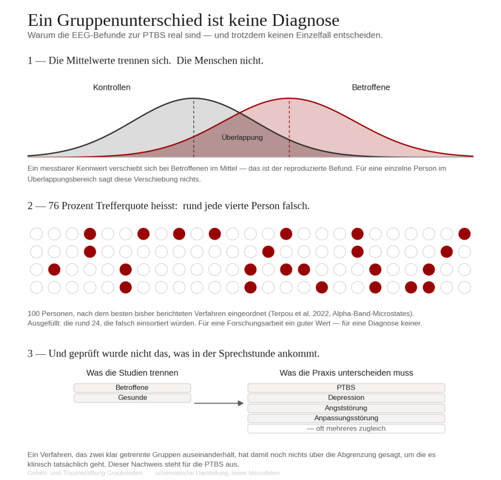
And yet: 76 per cent classification accuracy is a respectable value for a research study and entirely insufficient for a diagnosis. Roughly one person in four would be misclassified. The authors name the limitations openly: about half of the participants with PTSD were taking psychotropic medication, which affects the EEG; recording used a 19-channel system, which is comparatively coarse; and there was no independent test set on which the accuracy could have been confirmed — which, with small samples, regularly yields flattering figures. A promising finding, then, awaiting confirmation; not a finished tool.
The picture that emerges — and its holes
Put the findings together and a picture emerges of a nervous system on permanent alert: an altered resting pattern, altered stimulus processing, less autonomic flexibility. That fits the clinical experience of hyperarousal and watchfulness. It is plausible — and plausibility is dangerous, because it convinces before it is proven.
The review itself names the holes clearly. Meta-analyses of the spectral findings arrive at inconsistent results. The heterogeneity of PTSD and the differing methods prevent a uniform profile. The medication status of those examined is often unknown, although psychotropic drugs change the EEG. And the samples are mostly small — the Peddi study comprised 21 people, predominantly women, and only with partial PTSD symptoms. Such numbers allow a direction to be read off, not a diagnostic instrument.
In short: to this day there is no electrophysiological marker approved or validated for diagnosing PTSD in the individual case. Anyone claiming otherwise is confusing a group difference with a diagnosis.
The special case of neurofeedback
Because hyperarousal is so central, it is tempting to train the brain towards a calmer pattern through neurofeedback. There are studies and encouraging individual findings. The honest state of affairs, however, is that a protocol grounded in clear, reproduced target features is missing — the same problem that exists in autism. As long as it is not settled which measurable feature should be changed in which direction, neurofeedback in PTSD remains a promising field but not an established procedure. That distinction is part of being straight about it.
What is the measurement for, then?
If no marker makes the diagnosis — what does brain function analysis contribute? The answer is the same as in autism, and it is more modest and more robust than the promise of a «trauma test»: it offers a second, independent route to the same question.
Clinical diagnosis rests on observation and memory. A physiological finding arises by a different path. Where the two agree, the assessment becomes more robust. Where they diverge, that is not a contradiction but a pointer — to a comorbidity, a subtype, another explanation. Especially in a condition so often met with doubt, an additional route that does not depend on the telling is more than a technical gimmick.
And one more thing: a finding that can be measured can be repeated. That makes courses visible. Whether a treatment is moving anything could then be judged by something other than how it feels.
What the evidence does not support
The limits cannot be drawn clearly enough, because the temptation to make more of them is particularly strong in PTSD.
First, the available studies mostly distinguish people with PTSD from healthy controls — two clearly separated groups. Clinical practice looks different: there the task is to tell PTSD apart from depression, an anxiety disorder or an adjustment disorder, often with several of them present at once. A method that separates the affected from the healthy has therefore said nothing yet about what actually arrives in the consulting room.
Second, the findings are averaged over groups. A difference between the means of two groups says little about where a single person lies — the distributions almost always overlap.
Third, the heterogeneity of PTSD is not a marginal problem but the core of it. A hyperaroused and a dissociative course could show opposing EEG patterns. A single marker that captures both may not be a reasonable expectation at all.
And fourth, here as everywhere: a biomarker does not become valid by being plausible, but by being reproducible in independent data. For PTSD that proof is outstanding.
The December workshop places the four poles of ADHD, autism, PTSD and sensitivity side by side because they overlap in lived experience — in hyperarousal, in altered stimulus processing, in regulation knocked out of balance. PTSD shows in exemplary fashion what brain function analysis can be in this and what it cannot: no trauma detector, but a second look at a system that until now could almost only be narrated.
Sources
- Kovacevic, N.; Meghdadi, A.; Berka, C. Characterizing PTSD Using Electrophysiology: Towards A Precision Medicine Approach. Clinical EEG and Neuroscience, 2025, 56(4).
- Peddi, S. et al. Towards predicting posttraumatic stress symptom severity using portable EEG-derived biomarkers. Scientific Reports, 2025. Open Access.
- Terpou, B. A. et al. Spectral decomposition of EEG microstates in post-traumatic stress disorder. NeuroImage: Clinical, 2022. Open Access.